Cancer cells metabolism and acidity
Cellular energy metabolism is one of the main processes that is affected when a cell become malignant. Cancer cells take up excess glucose (even in the presence of oxygen) and use the glycolytic pathway more than normal cells do, a phenomenon known as the Warburg Effect or metabolic switch. As a result of glycolysis and overexpression of carbonic anhydrases the tumor became acidic. Moreover, due to the flux and the membrane potential, the extracellular pH is lowest at the surfaces of cancer cells and is significantly lower than normal physiological pH and the bulk extracellular pH in tumors. The low pH region persists at the cancer cell surface even in well-perfused tumor areas. Not only is acidity a hallmark of tumor development, but acidity also facilitates tumor growth. Cancer cells alter their metabolism to support their rapid proliferation and dissemination across the body, which represents a selective growth advantage, as cells become more motile and resistant to apoptosis. We study cancer cells metabolism and develop approaches to measure pH at the surface of cancer cells.
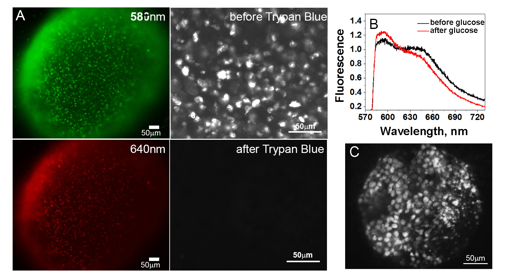 |
Images of tumor spheroids and Trypan Blue assay (A). Fluorescence images of HeLa tumor spheroids treated with the SNARF pHLIP peptide at pH 6.6 were acquired using 580 nm and 640 nm emission filters. The SNARF pHLIP peptide images of HeLa tumor spheroids are shown before and immediately after the addition of cell-impermeable Trypan Blue, which clearly demonstrates extracellular localization of SNARF fluorophore when pHLIP is inserted into plasma membrane of cells. pH measured at the surfaces of cancer cells in tumors in vivo (B). Fluorescence spectra recorded from tumors in live mice (skin is removed from the tumor site) 4 hours after administration of SNARF pHLIP peptide as a single tail vein injection before and after IP injection of 125 mg of glucose. pH imaging of cancer cells ex vivo (C). Fluorescence image obtained from a HeLa tumor specimen treated ex vivo with the SNARF pHLIP peptide in the presence of glucose, followed by washing. Images are from paper: Anderson et al, 2016, Proc Natl Acad Sci U S A. |